-
Про лікарню
![]() LISOD - онкологічна лікарня з повним циклом допомоги: профілактики, діагностики, лікування і реабілітації з використанням міжнародних стандартів на засадах доказової медицини. У LISOD використовується необхідне сучасне високотехнологічне обладнання для діагностики та лікування онкологічних захворювань.
Докладніше
LISOD - онкологічна лікарня з повним циклом допомоги: профілактики, діагностики, лікування і реабілітації з використанням міжнародних стандартів на засадах доказової медицини. У LISOD використовується необхідне сучасне високотехнологічне обладнання для діагностики та лікування онкологічних захворювань.
Докладніше
-
Наша команда
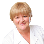
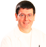
Алла Вінницька
![]()
Онкогінеколог. Доктор медичних наук. Медичний директор LISOD.
Член Європейського товариства медичних онкологів.
Онкогінеколог світового рівня. Проводить сучасну діагностику і лікування всіх гінекологічних захворювань: рак шийки матки, рак ендометрія, рак яєчників. -
Консультація
![Консультація ізраїльського онколога]()
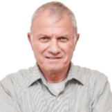
У LISOD ведуть прийом ізраїльські фахівці - професіонали з величезним досвідом роботи в кращих медичних центрах світу. Ізраїльські лікарі володіють методами лікування різних видів раку і надають ефективну допомогу при будь-яких онкологічних патологіях.
![Консультація онкогінеколога]()
Прийом веде Алла Вінницька - відомий онкогінеколог, доктор медичних наук, головний лікар LISOD. Алла Вінницька має багаторічний досвід профілактики, діагностики та максимально результативного лікування доброякісних і злоякісних захворювань жіночої статевої сфери.
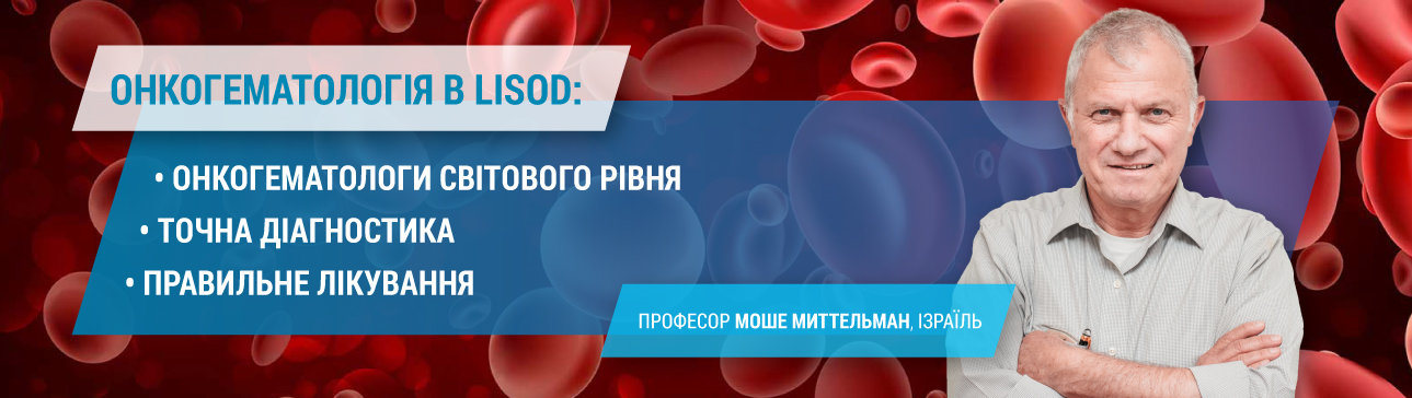
![Консультація онкогематолога]()
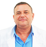
Консультацію проводять провідні ізраїльські та українські онкогематологи - експерти з багаторічним досвідом і знаннями в області лікування онкогематологічних захворювань. Пацієнтам доступна точна діагностика і ефективне лікування гематологічних захворювань, що відповідають всім сучасним вимогам і рекомендаціям міжнародних медичних протоколів (NCCN, AHA, EHA, ESMO).
![Консультація онкодерматолога]()
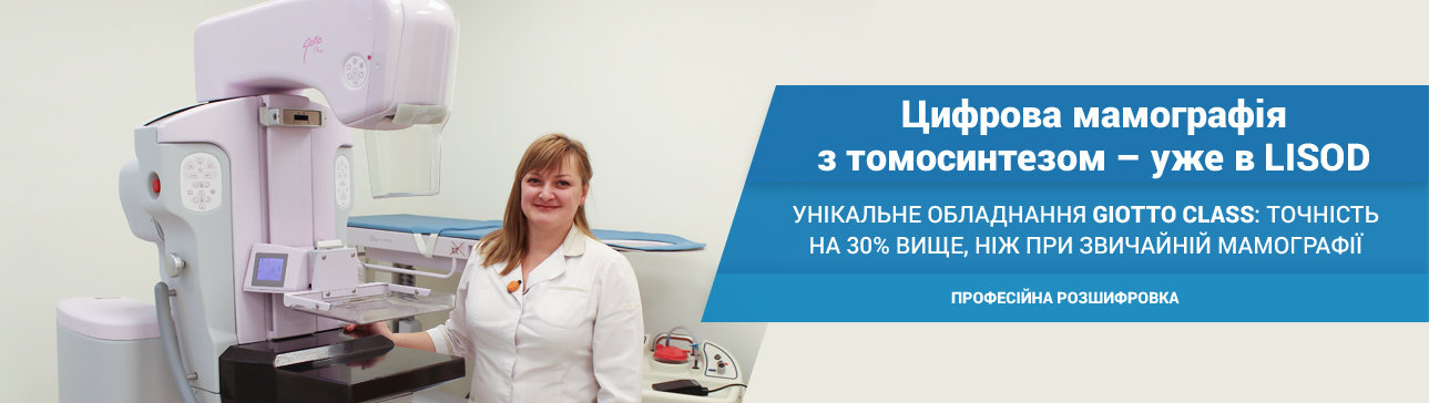
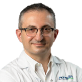
Консультацію проводять досвідчені онкодерматологи, експерти з діагностики та лікування захворювань шкіри. Пацієнти проходять обстеження на найсучаснішому обладнанні - FotoFinder's Bodystudio ATBM. Точна і швидка діагностика доброякісних і злоякісних новоутворень шкіри. Лікування злоякісних новоутворень шкіри відповідно до рекомендацій міжнародних медичних протоколів (NCCN, ESMO т.д.).
- Лікування раку
-
Попередь рак
-
Всі розділи
-
Про лікарню
![]() LISOD - онкологічна лікарня з повним циклом допомоги: профілактики, діагностики, лікування і реабілітації з використанням міжнародних стандартів на засадах доказової медицини. У LISOD використовується необхідне сучасне високотехнологічне обладнання для діагностики та лікування онкологічних захворювань.
LISOD - онкологічна лікарня з повним циклом допомоги: профілактики, діагностики, лікування і реабілітації з використанням міжнародних стандартів на засадах доказової медицини. У LISOD використовується необхідне сучасне високотехнологічне обладнання для діагностики та лікування онкологічних захворювань.
-
Наша команда
Алла Вінницька
![]()
Онкогінеколог. Доктор медичних наук. Медичний директор LISOD.
Член Європейського товариства медичних онкологів.
Онкогінеколог світового рівня. Проводить сучасну діагностику і лікування всіх гінекологічних захворювань: рак шийки матки, рак ендометрія, рак яєчників. -
Консультація
Консультація ізраїльського клінічного онколога в LISOD - це:
прийом фахівця зі світовим ім'ям, призначення тільки необхідних обстежень, консиліум лікарів різного профілю, розробка правильної тактики лікування, постійне курирування пацієнта.Клінічні онкологи LISOD спеціалізуються:
У LISOD ведуть прийом ізраїльські фахівці - професіонали з величезним досвідом роботи в кращих медичних центрах світу. Ізраїльські лікарі & nbsp; володіють методами лікування різних видів раку і нададуть ефективну допомогу при будь-онкологічної проблеми.
Прийом веде Алла Вінницька - відомий онкогінеколог, доктор медичних наук, професор, головний лікар LISOD. Алла Вінницька має багаторічний досвід профілактики, діагностики та максимально результативного лікування доброякісних і злоякісних захворювань жіночої статевої сфери.
- Підписатися на розсилку
- Записатися на прийом
-
Лікування раку
-
Попередь рак
-
Про лікарню
- Наші контакти
-
Обрати
лікаря -
Питання до LISOD
-
Записатися на прийом
 LISOD - онкологічна лікарня з повним циклом допомоги: профілактики, діагностики, лікування і реабілітації з використанням міжнародних стандартів на засадах доказової медицини. У LISOD використовується необхідне сучасне високотехнологічне обладнання для діагностики та лікування онкологічних захворювань.


 LISOD - онкологічна лікарня з повним циклом допомоги: профілактики, діагностики, лікування і реабілітації з використанням міжнародних стандартів на засадах доказової медицини. У LISOD використовується необхідне сучасне високотехнологічне обладнання для діагностики та лікування онкологічних захворювань.

Важливе про Lisod
Лікарня ізраїльської онкології в Україні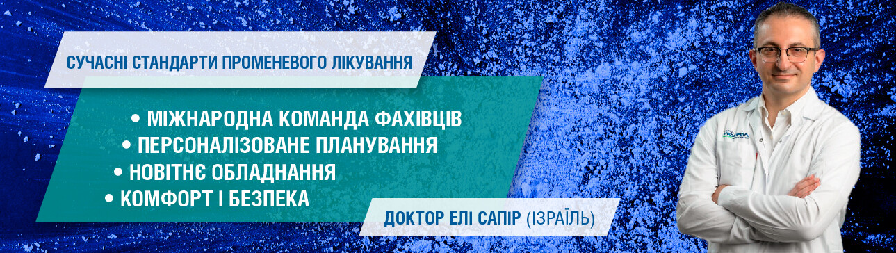
Ми лікуємо Комплексна діагностика і лікування раку в Україні
- Рак молочної залози
- Рак головного мозку
- Рак гортані
- Рак губи
- Рак дванадцятипалої кишки
- Рак шлунка
- Рак жовчного міхура
- Рак шкіри
- Рак легенів
- Лімфоми
- Рак матки
- Меланома
- Рак мигдалин
- Рак сечового міхура
- Рак печінки
- Рак стравоходу
- Рак підшлункової залози
- Рак статевого члена
Запис на прийом
ВИСОКОКЛАСНІ ІЗРАЇЛЬСЬКІ І УКРАЇНСЬКІ ЛІКАРІ


Чому LISOD?

Доказова медицина — застосування в щоденній практиці методів і технологій, ефективність яких доведена клінічними дослідженнями. Для кожного пацієнта LISOD консиліум фахівців розробляє оптимальний план діагностики і лікування, що відповідає рекомендаціям міжнародних протоколів. Призначаються тільки необхідні в конкретній клінічній ситуації процедури, водночас значно підвищується ймовірність одужання. Рівень лікування в LISOD відповідає рівню провідних онкологічних центрів Ізраїлю та Європи.
Ви можете бути впевнені, що фахівці Лікарні ізраїльської онкології володіють кращими сучасними методами лікування вашого захворювання.
Гістологічну верифікацію діагнозу LISOD проводить в патлабораторії Німеччини.

Пацієнт LISOD відразу потрапляє на прийом до ізраїльського клінічного онколога – фахівця з глибокими знаннями методів радіотерапії, хіміотерапії, гормональної терапії, з величезним досвідом діагностики та лікування різних видів раку. Завдяки цьому унікальному досвіду клінічний онколог встановлює діагноз, визначає ефективну тактику лікування. Такий підхід багато в чому забезпечує успіх у боротьбі з хворобою, що є вкрай важливим для кожного пацієнта.
Клінічні онкологи LISOD – авторитетні фахівці з Ізраїлю.

Ви отримуєте світовий рівень медичних послуг. В Україні лікуєтеся у зарубіжних фахівців – ізраїльських онкологів. Ви проходите діагностику та лікування з використанням сучасного обладнання. З вами поруч можуть перебувати рідні та близькі. У більшості випадків ви зберігаєте звичний спосіб життя. Економите кошти – вартість лікарських послуг в LISOD адаптована до умов України і набагато нижча, ніж вартість послуг такого ж рівня в інших європейських та ізраїльських клініках.
Лікарня ізраїльської онкології в Україні

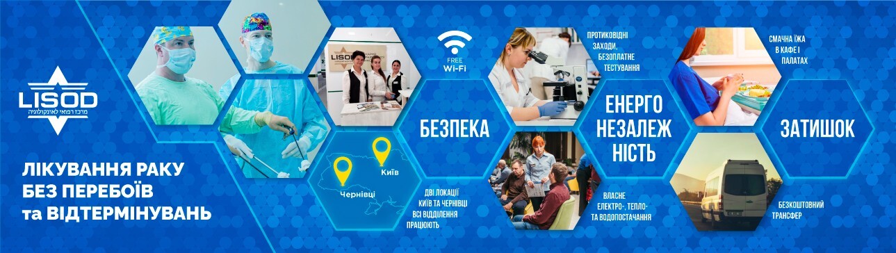
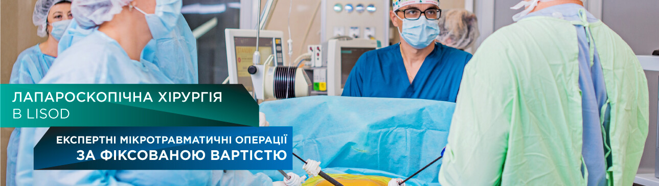
Пацієнтам LISOD доступні ефективні, перевірені, затверджені світовим медичним співтовариством методи лікування раку. Пацієнти проходять малотравматичне хірургічне лікування за допомогою лапароскопічного методу. Для кожного пацієнта використовують сучасні хіміотерапевтичні препарати згідно з протоколом. Завдяки глибоким знанням і досвіду онкологів променеве лікування проводиться максимально результативно з мінімальними побічними явищами.
Успіх у боротьбі з раком в LISOD досягнутий завдяки досвіду ізраїльських фахівців і застосуванню сучасного обладнання.

Для кожного пацієнта на самому початку лікування фахівці планують заходи щодо відновлення організму, розробляють індивідуальну програму реабілітації в декількох напрямках: медична та психологічна підтримка. Мета програми: забезпечити високу якість життя людини протягом всього медичного процесу; після закінчення лікування повернути пацієнта до того життя, яке він вів до хвороби.
Для пацієнтів LISOD послуги психолога, реабілітолога, фізіотерапевта і дієтолога безкоштовні.
Ваша думка важлива для нас

Спеціаліст з контролю якості обслуговування
Ваш коментар дозволить поліпшити якість обслуговування пацієнтів Лікарні ізраїльської онкології LISOD. Кожна думка допомагає нам постійно вдосконалювати рівень сервісу.
Гаряча лінія для відгуків:
Залиште свій відгук онлайн:
Медіа-центр


Наші страхові партнери


